|
Back to:
Blind Test XXXIII (ZEGWAN)
(Last updated February 2026)
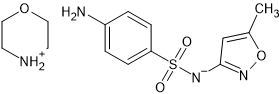
Figure 1. The molecular diagram of Blind Test XXXIII (ZEGWAN).
CSP studies
| REFCODE | |
| Formula | C4 H10 N1 O1 + . C10 H10 N3 O3 S1 - |
| Common Name | Blind Test XXXIIII |
| IUPAC Systematic Name | Morpholin-4-ium (4-aminophenyl)sulfonyl-(5-methylisoxazol-3-yl)azanide |
| Other Names | |
| CSD Refcodes | ZEGWAN, ZEGWAN01 |
| |
| Search Identifier | |
| Scientist | Rui Guo |
| Date | September 2021 |
| Publication | Hunnisett LM, et al, Acta Cryst B, 2024, 80(6), 517-547 |
| Energy model | |
| Study_ID | 10 |
| Programs | CrystalPredictor (2.4.3.2), CrystalOptimizer (2.4.8), DMACRYS (2.3.1.1) |
| Location on S Drive | CHEMISTRY_CPOSS\BlindTest2020-2022\XXXIII\CrystOpt1\ |
| Potential Description | CrystalOptimizer PBE1PBE/6-31+G(d,p) intramolecular + GDMA2.2(PCM,E=11(PBE1PBE/6-31+G(d,p))) DMA + FIT + Scheraga SO2 parameters |
| Intramolecular Description | PCM11(PBE1PBE/6-31+G(d,p)) |
| |
| Search Identifier | |
| Scientist | Louise Price, Rui Guo |
| Date | June 2022 |
| Publication | Hunnisett LM, et al, Acta Cryst B, 2024, 80(6), 548-574 |
| Energy model | |
| Study_ID | 11 |
| Programs | CCDC supplied structures, CrystalOptimizer (2.4.8), DMACRYS (2.3.1.1) |
| Location on S Drive | CHEMISTRY_CPOSS\BlindTest2020-2022\XXXIII\CrystOpt2\ |
| Potential Description | CrystalOptimizer PBE1PBE/6-31+G(d,p) intramolecular + GDMA2.2(PCM,E=11(PBE1PBE/6-31+G(d,p))) DMA + FIT + Scheraga SO2 parameters |
| Intramolecular Description | PCM11(PBE1PBE/6-31+G(d,p)) |
| pDFT structures | |
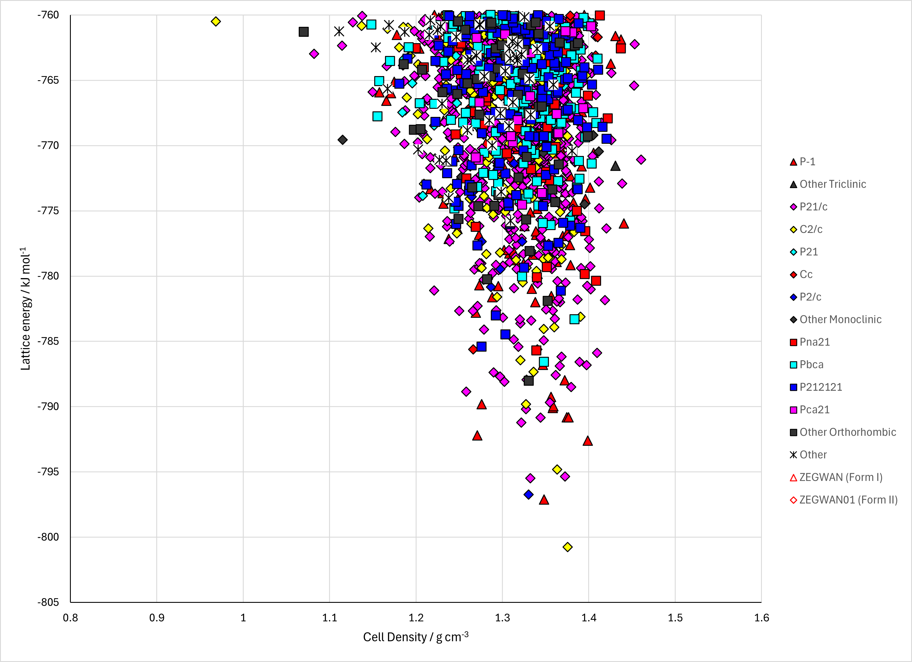
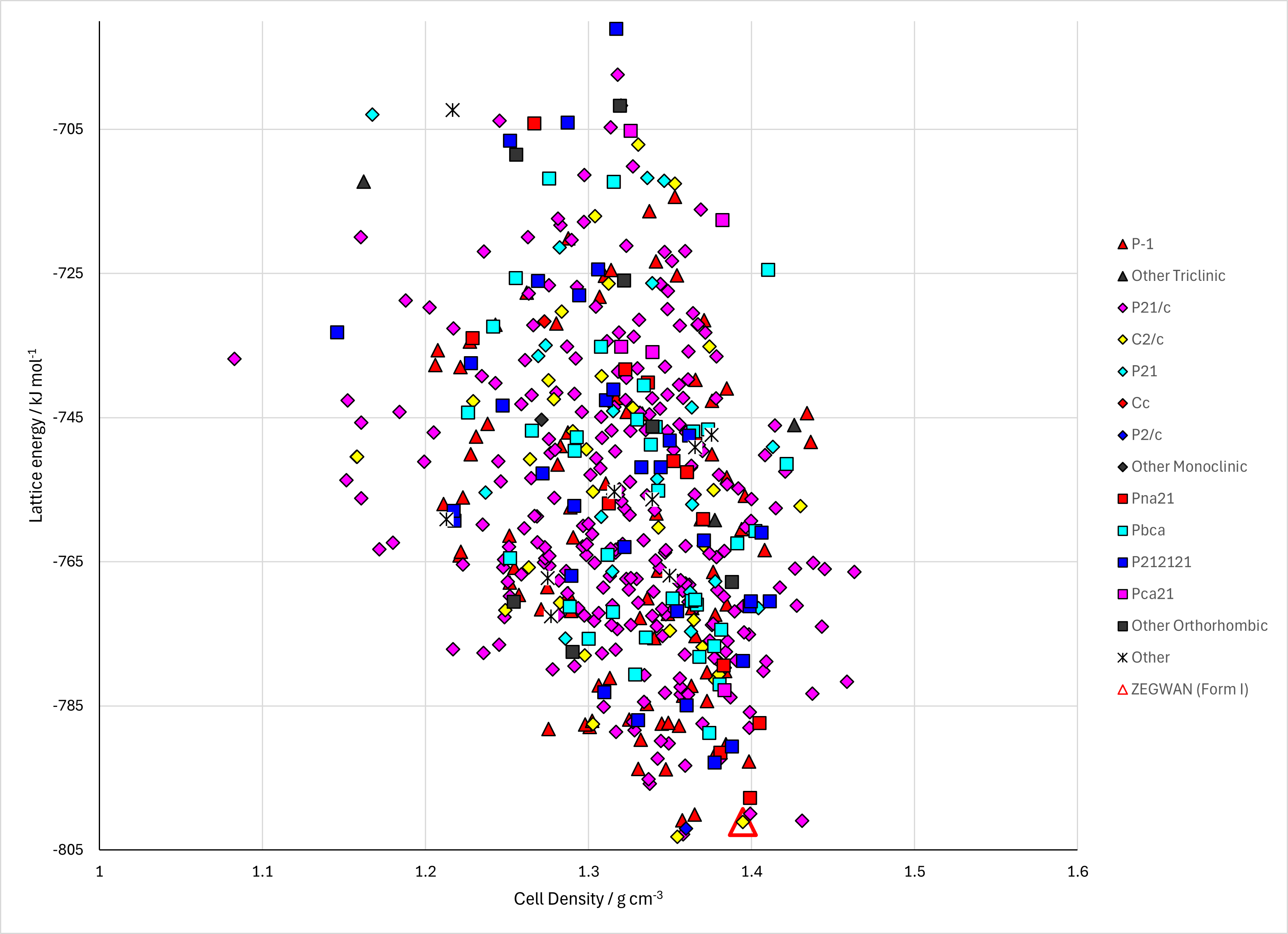
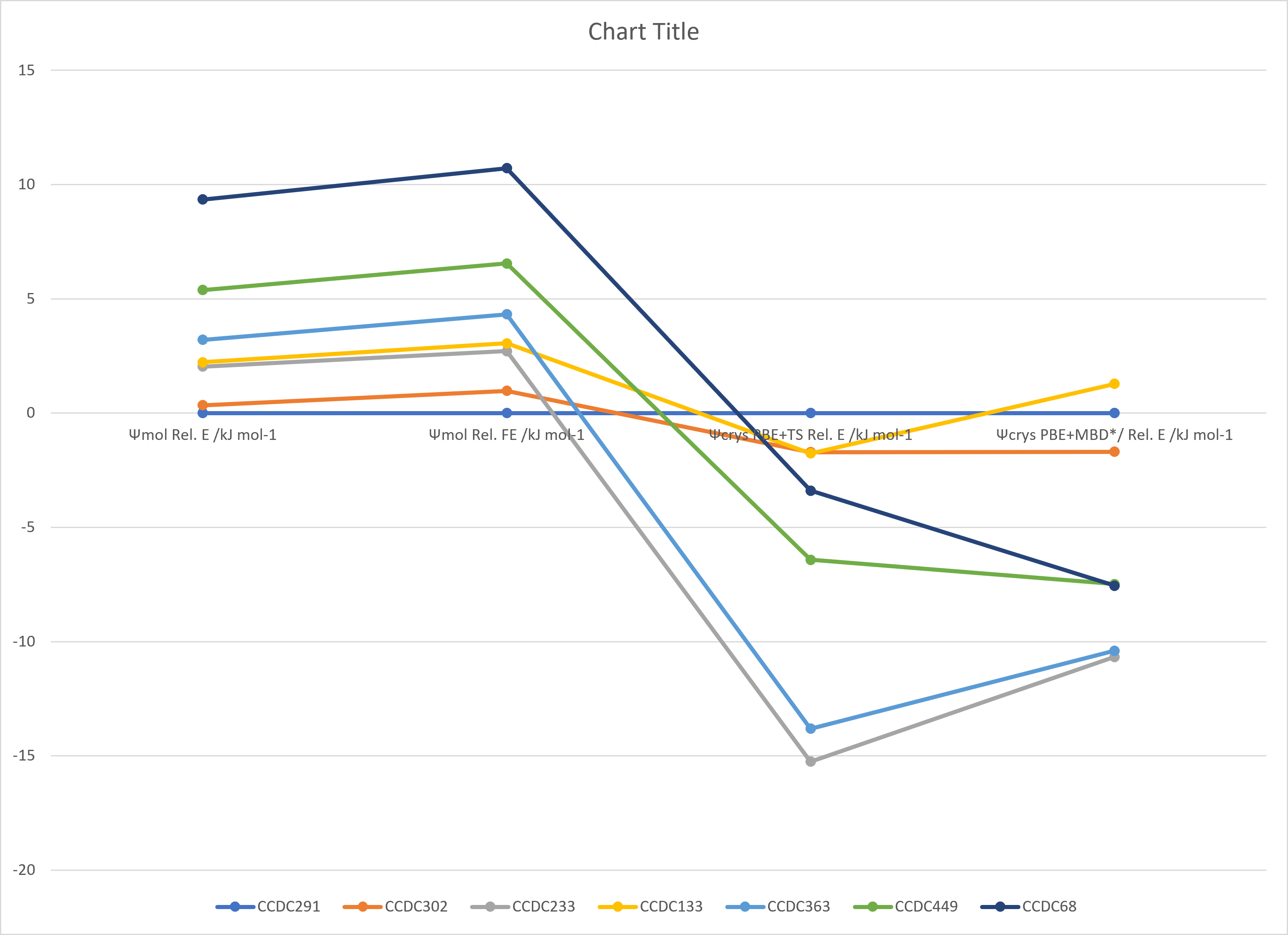
Figure 2. Lattice Energy Landscapes of Blind Test XXXIII (ZEGWAN). Energy models A1 and B1. Line chart comparing DFT+D energy models.
CSD structures (CSD version 6.01)
Table 1. Crystallographic information and experimental information of CSD entries for Blind Test XXXIII (ZEGWAN). Different polymorphs, as ascertained by structure matching of 20 molecule coordination sphere in Mercury, are coloured differently. Reference polymorphs are outlined in bold.
| REFCODE | Spacegroup | Z' | a / Å | b / Å | c / Å | α / ° | β / ° | γ / ° | density / g cm-3 | Form | R factor | T / K | Year | Comment / reference | REFCODE |
|---|
| ZEGWAN | C2/c | 1.0 | 24.9972 | 10.5875 | 12.9323 | 90.0 | 104.502 | 90.0 | 1.3646 | Form I polymorph | 3.58 | 296.5 K | 2022 | None | ZEGWAN |
| ZEGWAN01 | Pna21 | 1.0 | 11.7407 | 12.4207 | 11.3775 | 90.0 | 90.0 | 90.0 | 1.3627 | Form II polymorph | 2.32 | 297.1 K | 2022 | None | ZEGWAN01 |
|