(Last updated December 2025)
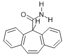
Figure . The molecular diagram of Cytenamide.
| REFCODE | SODNOP |
|---|---|
| Formula | C16 H13 N1 O1 |
| Common Name | Cytenamide |
| IUPAC Systematic Name | 5H-dibenzo(a,d)cycloheptatriene-5-carboxamide |
| Other Names | |
| CSD Refcodes | SOGLEG, SODNOP |
| Search Identifier | A |
| Scientist | Rui Guo |
| Date | 2016 |
| Publication | Rui is working on it |
| Energy model | 1 |
| Study_ID | 10 |
| Programs | Flexible CrystalPredictor (2.1.01), CrystalOptimizer (2.4.m), DMACRYS (2.2.0.1) |
| Location on S Drive | /CHEMISTRY_CPOSS/CarbamazepineSeries/Cytenamide_CO |
| Potential Description | CrystalOptimizer with PBE0/6-31G(d,p) Intra and GDMA2.2(PBE0/6-31G(d,p)) + FIT |
| Energy model | 2 |
| Study_ID | 30 |
| Programs | Study_ID=10, DMACRYS (2.2.0.1) |
| Location on S Drive | /CHEMISTRY_CPOSS/CarbamazepineSeries/Cytenamide_PCM |
| Potential Description | GDMA2.2(PCMdielectric3(PBE0/6-31+G(d))) + FIT |
| Search Identifier | B |
| Scientist | Louise Price |
| Date | 2010 |
| Publication | No publication planned |
| Energy model | 1 |
| Study_ID | 0 |
| Programs | MOLPAK, DMAREL (4.1.1) |
| Location on S Drive | \\CHEMISTRY_CPOSS\\0-EarlySearches\\home\\louise_price.eminerals\\cytenamide |
| Potential Description | DMA + FIT |
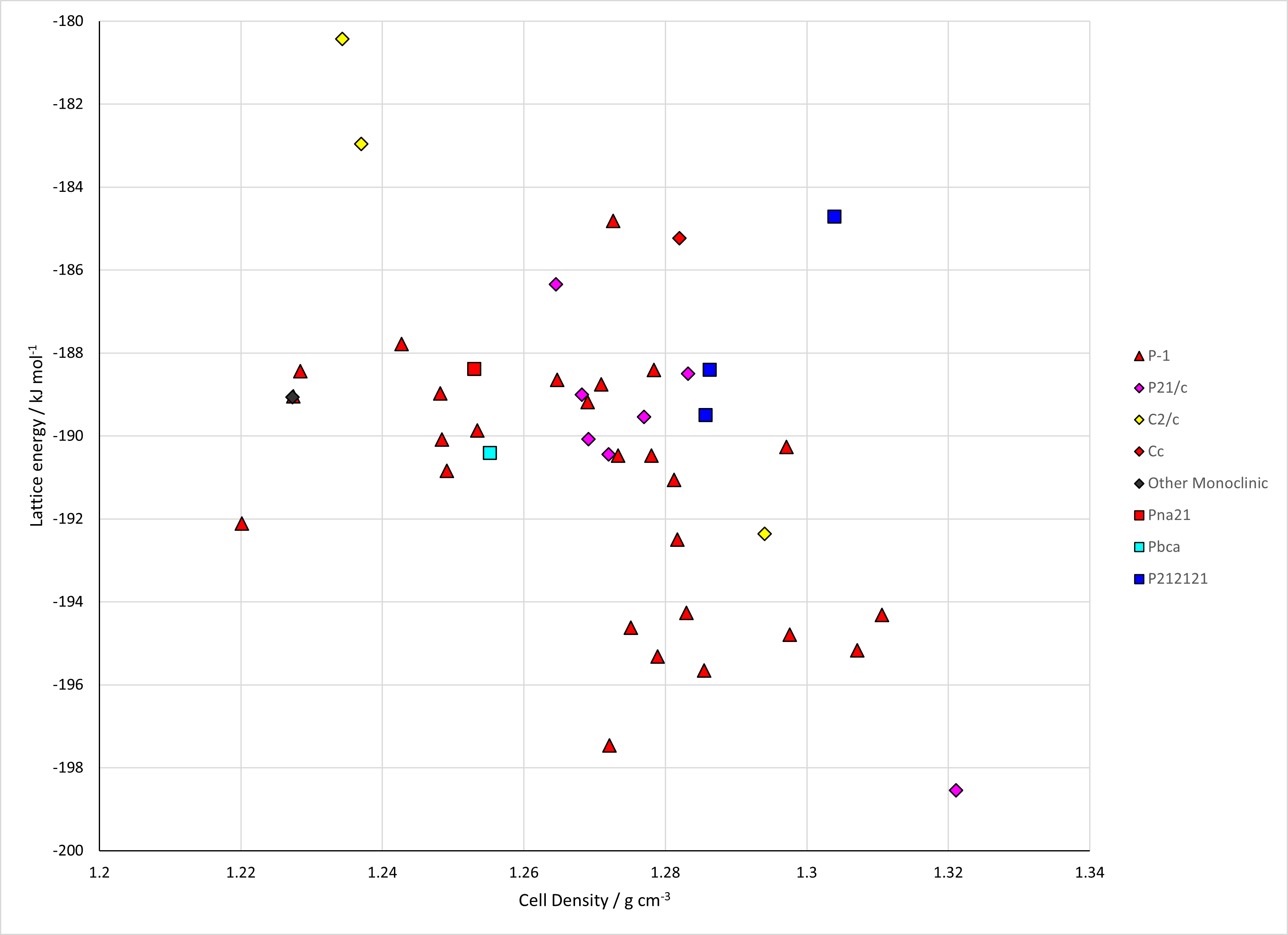
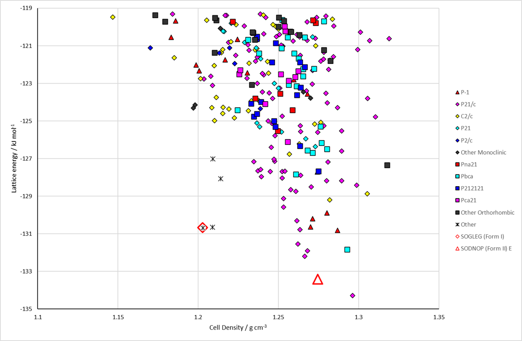
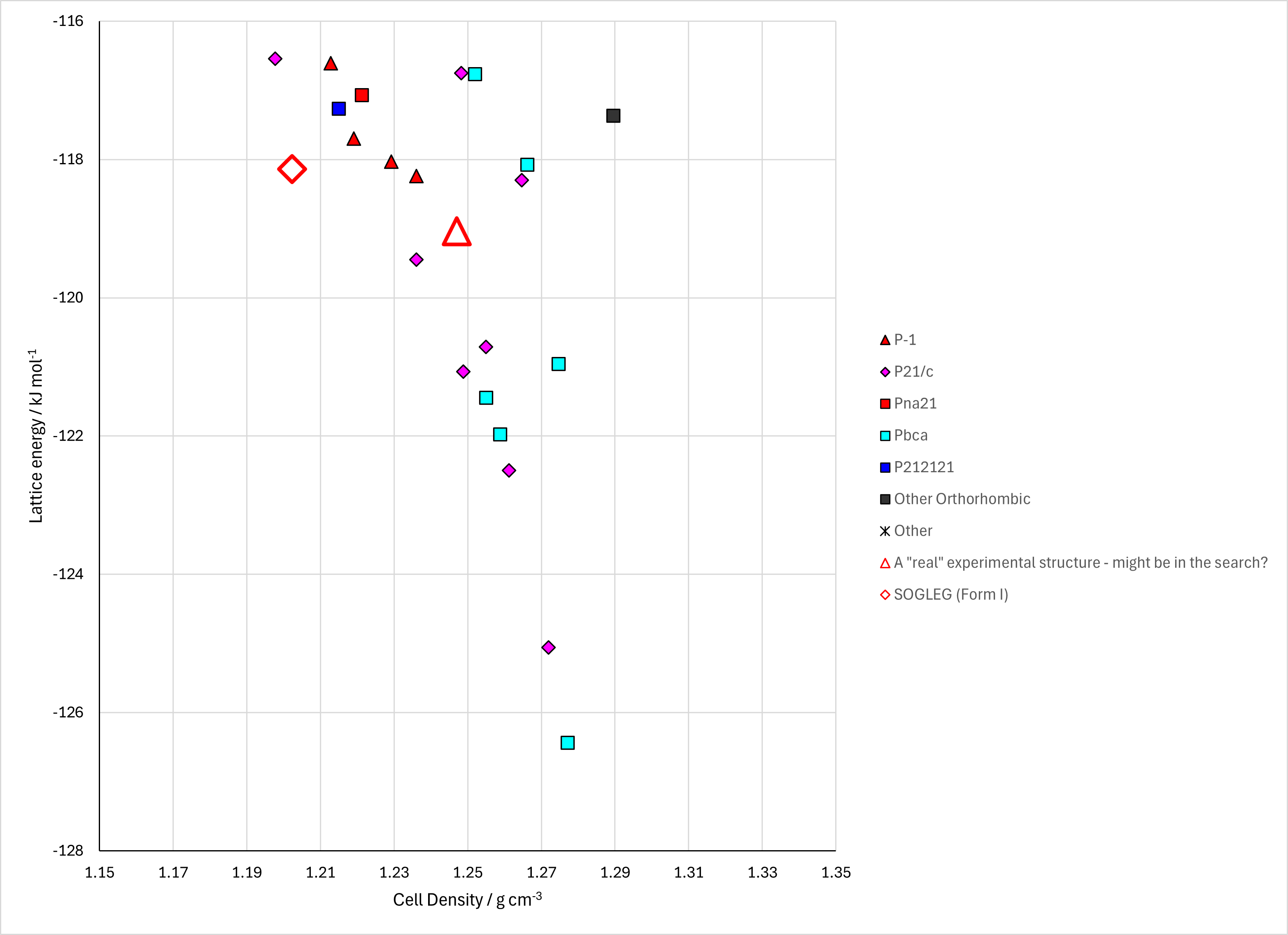
Figure . (Top) crystal energy landscape of Cytenamide from Study_ID=10. (Bottom left) crystal energy landscape of Cytenamide from Study_ID=0. (Bottom right) relative energies by method of key structures.
Table . Crystallographic information for CSD entries for Cytenamide. Different polymorphs are coloured differently.
| REFCODE | space group | Z’ | a / Å | b / Å | c / Å | α / ° | β / ° | γ / ° | density / g cm-3 | Form |
|---|---|---|---|---|---|---|---|---|---|---|
| SODNOP | P-1 | 4 | 5.810(<1) | 19.632(<1) | 21.709(<1) | 85.92(<1) | 86.16(<1) | 84.48(<1) | 1.274 | II |
| SOGLEG | R-3 | 1 | 33.908(1) | 33.908(1) | 5.675(<1) | 90 | 90 | 120 | 1.244 | I |
Table . Experimental information for CSD entries for Cytenamide.
| REFCODE | space group | R factor | T / K | Year | Comments |
|---|---|---|---|---|---|
| SODNOP | P-1 | ? | 293 | 2008 | From powder. The authors refer to this as Cytenamide form II, but the structure they refer to as Cytenamide form I, in the same paper, has residual solvent molecules in the lattice (accounted for using SQUEEZE) and thus cannot be a true polymorph of the form reported here.1 Heating form I to 498 K and cooling to RT. |
| SOGLEG | R-3 | 7.1 | 120 | 2008 | Residual peaks attributable to disordered solvent molecules have been accounted for using the SQUEEZE procedure, so the structure was refined as an unsolvated model. The authors refer to this as Cytenamide form I, but it has residual solvent molecules in the lattice (accounted for using SQUEEZE) and thus cannot be a true polymorph of the form II structure also reported in the paper.1 Crystallized from industrial methylated spirits. |
Form I (SOGLEG) is isostructural with carbamazepine form II.
Form II (SODNOP) is isostructural with carbamazepine form I and cyheptamide form II.
Left: the packing of CYT molecules in form I (R-3, Z′ = 1), viewed along the c-axis.15 The colour scheme applied to the molecules in the lower section marks one example of the common 2D structure present in both forms of CYT. Right: the packing of CYT molecules in form II (P-1, Z′ = 4), viewed along the a-axis. The four crystallographically independent stacks of molecules are drawn in different colours.
1. A. J. Florence, C. T. Bedford, F. P. A. Fabbiani, K. Shankland, T. Gelbrich, M. B. Hursthouse, N. Shankland, A. Johnston and P. Fernandes, CrystEngComm, 2008, 10, 811-813.