
Back to:
| Ordered alphabetically by REFCODE | Ordered alphabetically by Common name | Ordered by molecular formula | Grouped by family |
Fenamic Acid
(Last updated November 2024)
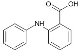
Figure 1. The molecular diagram of Fenamic Acid.
CSP studies
This system was studied by Ogaga Uzoh, as part of his PhD. He ran a search in CrystalPredictor, followed by CrystalOptimizer refinement and PCM refinement. At the moment, I don’t know which versions of CrystalPredictor or CrystalOptimizer were used, or whether the search was rigid or flexible. Looking at the summary.out files, the variation in starting values of dih9 and dih17 is very small, but they are all different values. Therefore, it looks like a flexible search. Similarly, the variation in bnd2 is small, so it appears to have been a CrystalPredictor2 search (CrystalPredictor1 would have this bond within a rigid fragment being a constant). It’s definitely CrystalOptimizer refinement, although the gas phase energy of opt was not included in the mol.input file.
Very few structures were optimized with CrystalOptimizer, and even fewer had the PCM refinement added. A related search by Ogaga had to be repeated later for exactly this reason – in that case one of the experimental crystal structures was missed as it was slightly too high on the CrystalPredictor energy ranking.
While compiling the structures and information for the energy comparison paper, it was noticed that different angles were optimized with CrystalOptimizer for each of the molecules in this family. Also, structurally equivalent packings were to be included for the whole family of molecules. This necessitated reoptimization with CrystalOptimizer prior to optimization with CASTEP.
| REFCODE |
| ||
| Formula | C13 H12 N1 O2 | ||
| Common Name | 2-Aniliobenzoic Acid | ||
| IUPAC Systematic Name | 2-(phenylamino)benzoic acid | ||
| Other Names | N-phenyl-anthranilic acid | ||
| CSD Refcodes | QQQBTY02 | ||
| Search Identifier |
| ||
| Scientist | Ogaga Uzoh | ||
| Date | 2012 | ||
| Publication | Uzoh OG, Cruz-Cabeza AJ, Price SL 2012. Cryst Growth Des 12, 4230-4239. | ||
| Energy model |
| ||
| Study_ID | 30 | ||
| Programs | Study_ID=10, DMACRYS (2.0.8) | ||
| Location on S Drive | /CHEMISTRY_CPOSS/Fenamates/FenamicAcid/QQQBTY_PCM | ||
| Potential Description | GDMA2.2(PCMdielectric3(PBE1PBE/6-31+G(d))) + FIT | ||
| Intramolecular Description | PBE1PBE/6-31+G(d) | ||
| Energy model |
| ||
| Study_ID | 10 | ||
| Programs | Flexible CrystalPredictor (1.6), dmaflex-Quick, CrystalOptimizer (2.2), DMACRYS (2.0.4) | ||
| Location on S Drive | /CHEMISTRY_CPOSS/Fenamates/FenamicAcid/QQQBTY_CO | ||
| Potential Description | CrystalOptimizer PBE1PBE/6-31+G(d) intramolecular energy with PBE1PBE/6-31+G(d) charge density and FIT potential | ||
| Intramolecular Description | PBE1PBE/6-31+G(d) | ||
| Energy model |
| ||
| Study_ID | 11 | ||
| Programs | Study_ID=10, CrystalOptimizer (2.4.7), DMACRYS (2.3.1.1) | ||
| Location on S Drive | /CHEMISTRY_CPOSS/Fenamates/FenamicAcid/QQQBTY_DFT | ||
| Potential Description | CrystalOptimizer PBE1PBE/6-31+G(d) intramolecular energy with PBE1PBE/6-31+G(d) charge density and FIT potential | ||
| Intramolecular Description | PBE0/6-31+G(d) | ||
| pDFT structures |
| ||
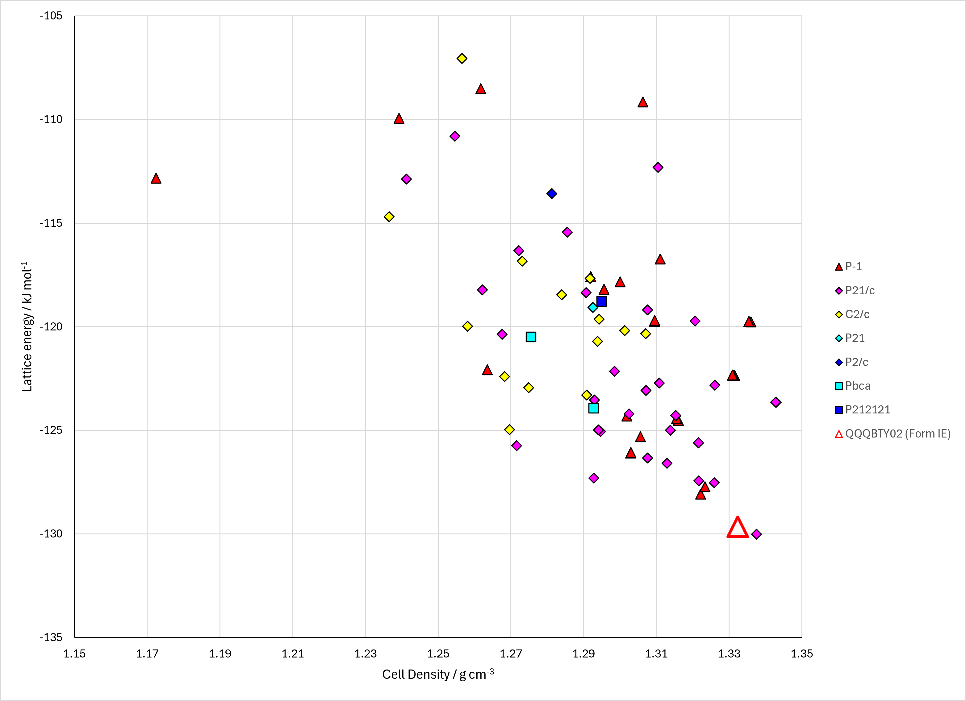
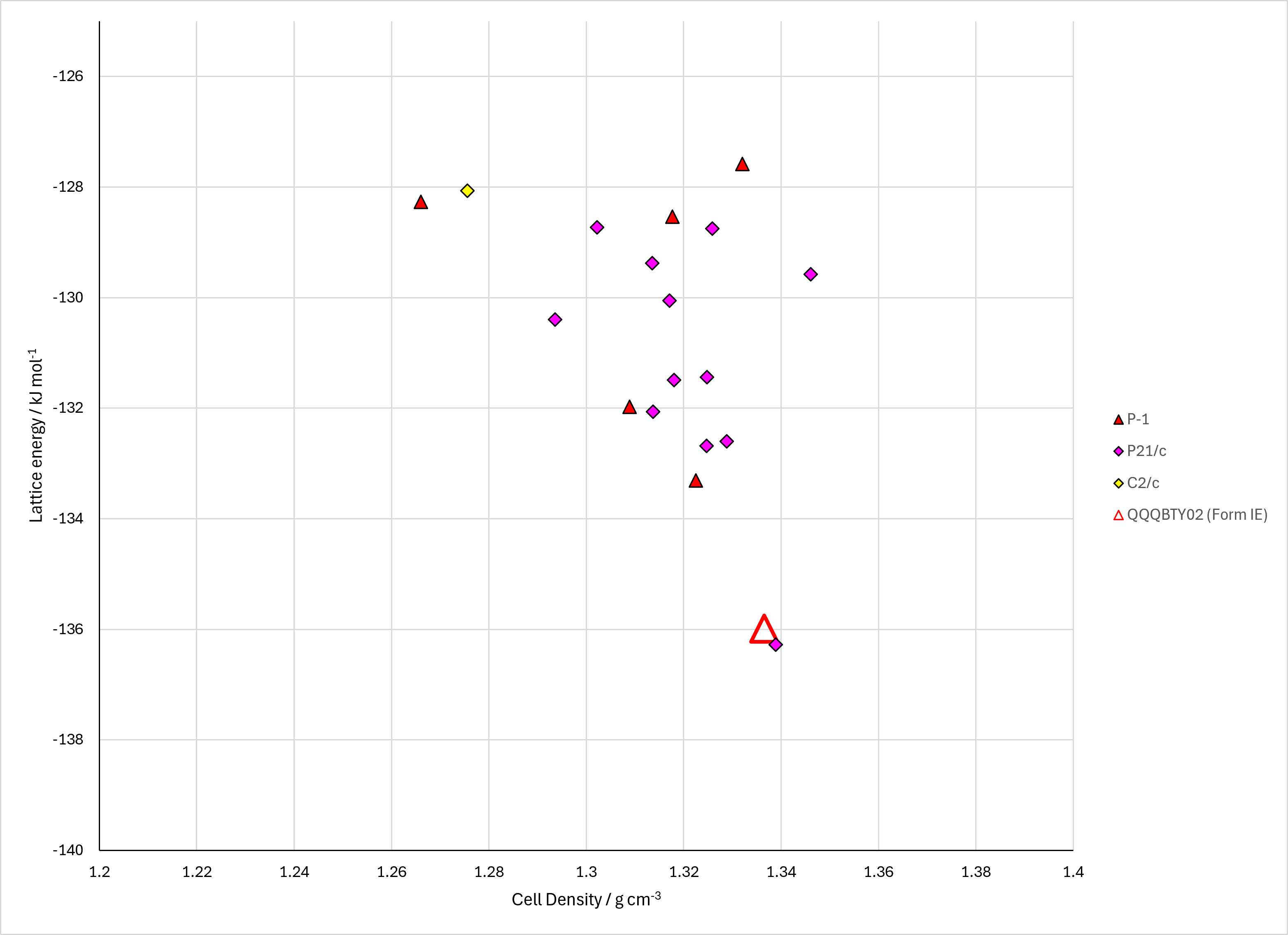
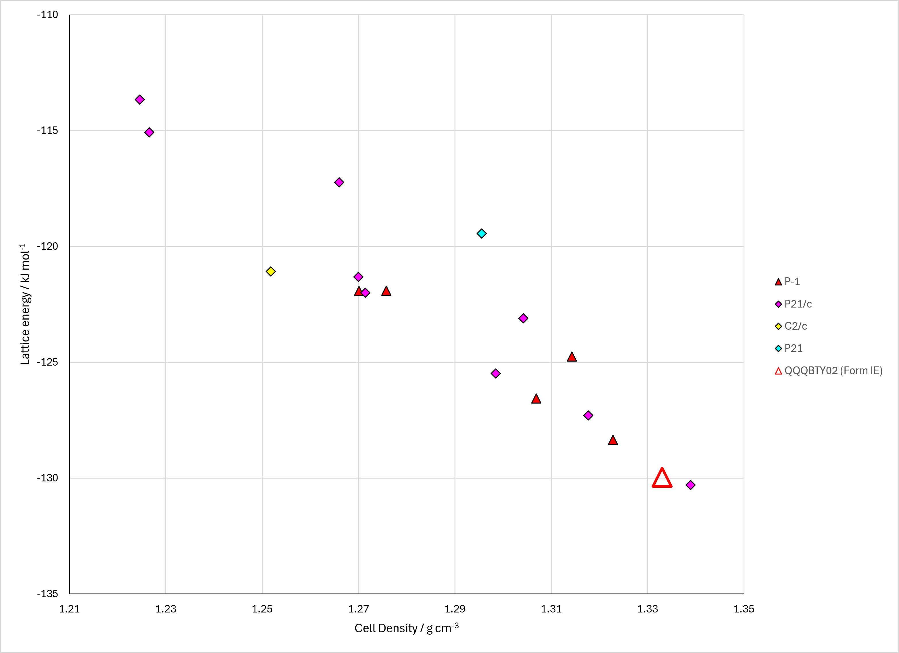
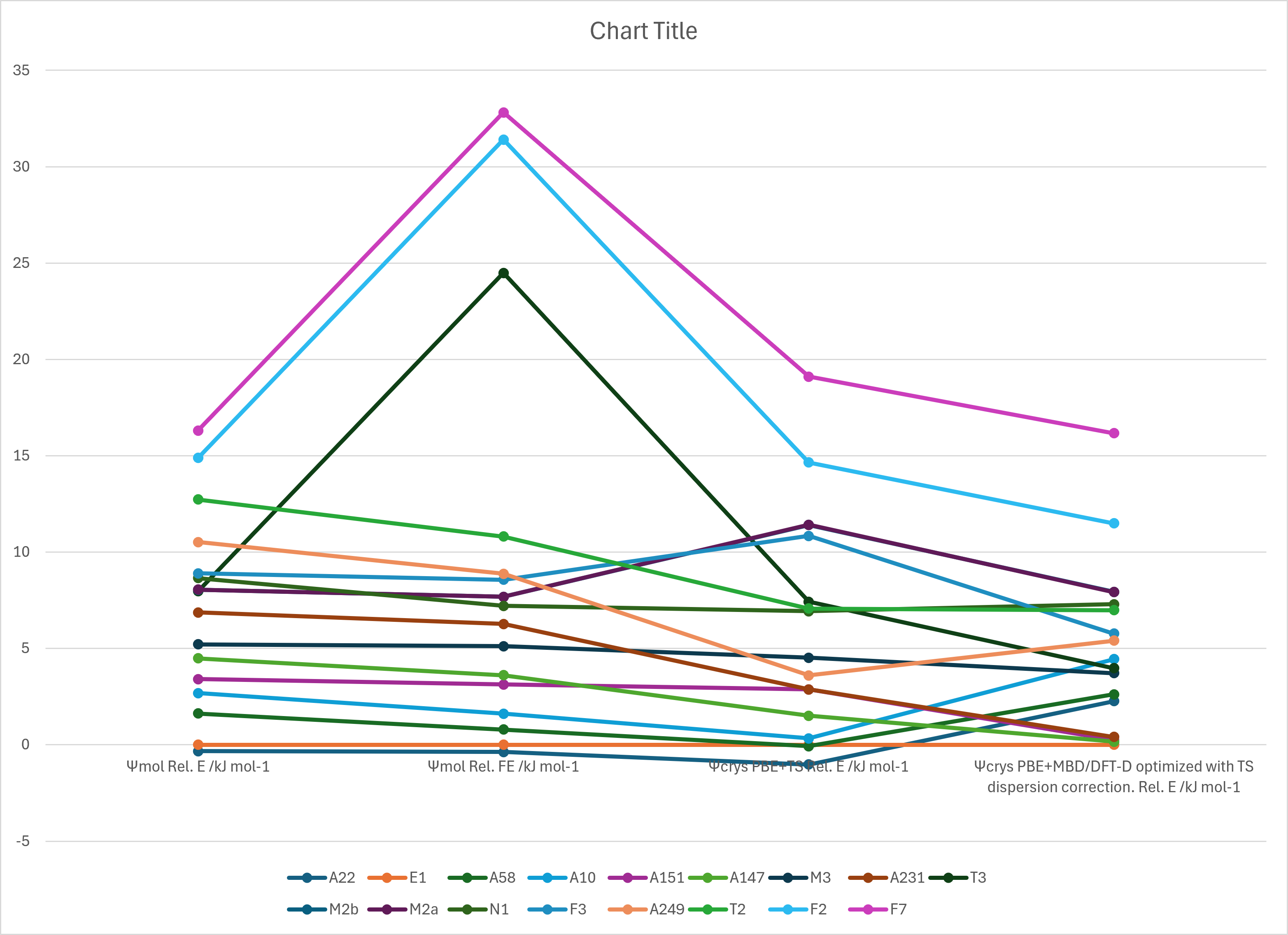
Figure 2. Lattice Energy Landscapes of Fenamic Acid. Energy models A1, A2 and A3. Line chart comparing DFT+D energy models.
CSD structures (CSD version 5.43 with Mar Jun Sep and Nov 2022 updates)
Table 1. Crystallographic information and experimental information of CSD entries for Fenamic Acid. Different polymorphs, as ascertained by structure matching of 20 molecule coordination sphere in Mercury, are coloured differently. Reference polymorphs are outlined in bold.
| REFCODE | Spacegroup | Z' | a / Å | b / Å | c / Å | α / ° | β / ° | γ / ° | density / g cm-3 | Form | R factor | T / K | Year | Comment / reference | REFCODE |
|---|---|---|---|---|---|---|---|---|---|---|---|---|---|---|---|
| QQQBTY | P-1 | 2.0 | 9.83 | 14.1200 | 8.03 | 90.05 | 106.60000 | 91.600000 | 0.0 | None | None | None | 1969 | None | QQQBTY |
| QQQBTY01 | P-1 | 2.0 | 8.0995 | 9.8268 | 14.0593 | 85.960000 | 88.62 | 73.39 | 1.3241 | None | 7.4 | None | 1987 | 10.1107/S0108270187093557 | QQQBTY01 |
| QQQBTY02 | P-1 | 2.0 | 8.08 | 9.812 | 14.0430 | 85.960000 | 88.640000 | 73.45 | 1.3304 | None | 4.89 | at 293 K | 2009 | 10.1039/b900476a | QQQBTY02 |
The Word Document has some crystallization methods.
Other Notes
The original study indicated that structure 22 was a reasonable approximation to the experimental Z’=2 structure. Louise thinks that structures 2 and 58 are closer. For the time being, nothing is marked as the experimental structure apart from the Z’=2 structure. The Word Document shows as overlay.
Of the 77 CSP-generated crystal structures on the CrystalOptimizer landscape, all have R_2^2 (8) hydrogen bonding apart from structures 249, 352, 415, 450, which have C_1^1 (4) hydrogen bonding, and structure 5675 which has R_2^2 (12) hydrogen bonding (NH···O=C).
© UCL Chemistry Department 2022. This page was last updated on 30 Sep 2026. If you have any problems with this page please email the WebMaster