
Back to:
| Ordered alphabetically by REFCODE | Ordered alphabetically by Common name | Ordered by molecular formula | Grouped by family |
5-Cyanouracil
(Last updated January 2026)
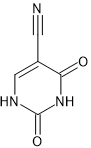
Figure 1. The molecular diagram of 5-Cyanouracil.
CSP studies
| REFCODE |
| ||
| Formula | C5 H3 N3 O2 | ||
| Common Name | 5-cyanouracil | ||
| IUPAC Systematic Name | 5-cyanouracil | ||
| Other Names | 5-cyanouracil | ||
| CSD Refcodes | BOLLOE | ||
| Search Identifier |
| ||
| Scientist | Nizar Issa | ||
| Date | 2005-7 | ||
| Publication | Barnett SA, Hulme AT, Issa N, Lewis TC, Price LS, Tocher DA, Price SL 2008. New J Chem 32 1761-1775. | ||
| Energy model |
| ||
| Study_ID | 0 | ||
| Programs | MOLPAK, DMAREL (4.1.1) | ||
| Location on S Drive | \CHEMISTRY_CPOSS\0-EarlySearches\home\louise_price.eminerals\5-cyanouracil | ||
| Potential Description | DMA + FIT | ||
| Intramolecular Description | MP2/6-31G(d,p) | ||
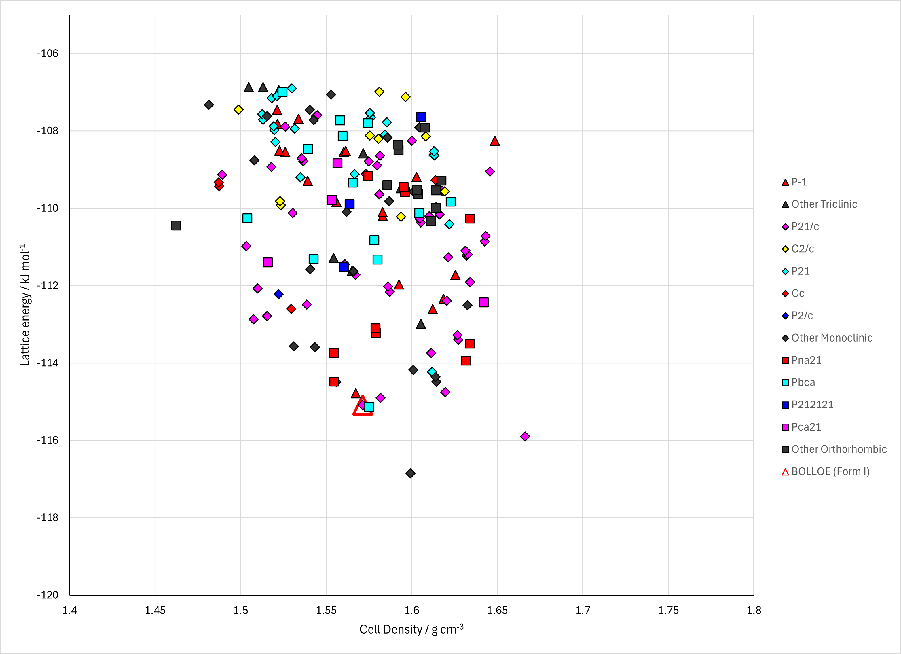
Figure 2. Lattice Energy Landscape of 5-Cyanouracil. Energy model A1.
CSD structures (CSD version 6.01)
Table 1. Crystallographic information and experimental information of CSD entries for 5-Cyanouracil. Different polymorphs, as ascertained by structure matching of 20 molecule coordination sphere in Mercury, are coloured differently. Reference polymorphs are outlined in bold.
| REFCODE | Spacegroup | Z' | a / Å | b / Å | c / Å | α / ° | β / ° | γ / ° | density / g cm-3 | Form | R factor | T / K | Year | Comment / reference | REFCODE |
|---|---|---|---|---|---|---|---|---|---|---|---|---|---|---|---|
| BOLLOE | P21/n | 1.0 | 9.0098 | 6.6035 | 9.1805 | 90.0 | 98.124 | 90.0 | 1.6840 | None | 3.98 | at 150 K | 2008 | 10.1039/b806763e | BOLLOE |
© UCL Chemistry Department 2022. This page was last updated on 30 Sep 2026. If you have any problems with this page please email the WebMaster