
Back to:
| Ordered alphabetically by REFCODE | Ordered alphabetically by Common name | Ordered by molecular formula | Grouped by family |
Tetrazol Pyrimidine
(Last updated April 2026)
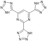
Figure 1. The molecular diagram of Tetrazol Pyrimidine.
CSP studies
| REFCODE |
| ||
| Formula | C7 N14 H4 | ||
| Common Name | Tetrazol-pyrimidine | ||
| IUPAC Systematic Name | 2,4,6-tri(1H-tetrazol-1-yl)pyrimidine | ||
| Other Names | |||
| CSD Refcodes | |||
| Search Identifier |
| ||
| Scientist | Luca Iuzzolino | ||
| Date | 2016 | ||
| Publication | No publication planned. | ||
| Energy model |
| ||
| Study_ID | 30 | ||
| Programs | Study_ID=10, DMACRYS (2.2.0.1) | ||
| Location on S Drive | \CHEMISTRY_CPOSS\Tetrazol\Tetrazol-pyrimidine\PCM | ||
| Potential Description | GDMA2.2(PCMdielectric3(PBE0/6-31G(d,p)) + FIT | ||
| Intramolecular Description | PCM3(PBE1PBE/6-31G(d,p)) | ||
| Energy model |
| ||
| Study_ID | 10 | ||
| Programs | CrystalPredictor, CrystalOptimizer, DMACRYS (2.2.0.1) | ||
| Location on S Drive | \CHEMISTRY_CPOSS\Tetrazol\Tetrazol-pyrimidine\CrystOpt | ||
| Potential Description | CrystalOptimizer with GDMA2.2(PBE1PBE/6-31G(d,p)) + FIT | ||
| Intramolecular Description | PBE1PBE/6-31G(d,p) | ||
| Energy model |
| ||
| Study_ID | 11 | ||
| Programs | Study_ID=10, CrystalOptimizer, DMACRYS (2.2.0.1) | ||
| Location on S Drive | \CHEMISTRY_CPOSS\Tetrazol\Tetrazol-pyrimidine\CrystOpt_will | ||
| Potential Description | CrystalOptimizer with GDMA2.2(PBE1PBE/6-31G(d,p)) + Will | ||
| Intramolecular Description | PBE1PBE/6-31G(d,p) | ||
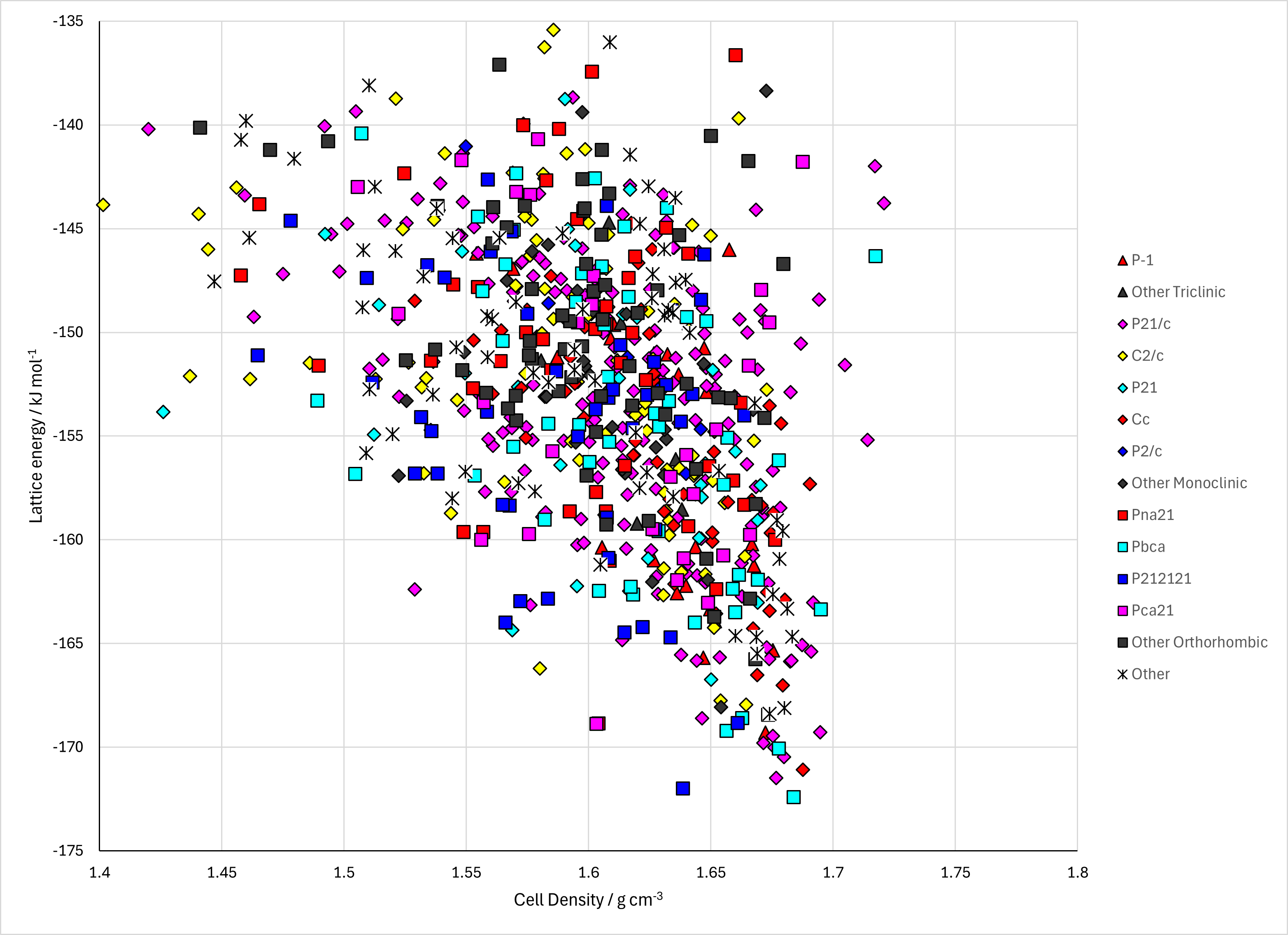
Figure 2. Lattice Energy Landscape of Tetrazol Pyrimidine. Energy model A3.
CSD structures (CSD version 6.01 with Feb 2026 update)
Table 1. Crystallographic information and experimental information of CSD entries for Tetrazol Pyrimidine. Different polymorphs, as ascertained by structure matching of 20 molecule coordination sphere in Mercury, are coloured differently. Reference polymorphs are outlined in bold.
| REFCODE | Spacegroup | Z' | a / Å | b / Å | c / Å | α / ° | β / ° | γ / ° | density / g cm-3 | Form | R factor | T / K | Year | Comment / reference | REFCODE |
|---|
© UCL Chemistry Department 2022. This page was last updated on 30 Sep 2026. If you have any problems with this page please email the WebMaster