|
Back to:
Uric Acid
(Last updated January 2026)
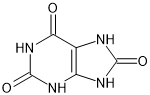
Figure 1. The molecular diagram of Uric Acid.
CSP studies
| REFCODE | |
| Formula | C5 H4 N4 O3 |
| Common Name | Uric acid |
| IUPAC Systematic Name | 7,9-dihydro-1H-purine-2,6,8(3H)-trione |
| Other Names | 2,6,8-trioxypurine, 7,9-dihydro-1H-purine-2,6,8(3H)-trione, Lithic acid, Purine-2,6,8(1H,3H,9H)-trione, 1H-Purine-2,6,8-triol, 2,6,8-Trioxopurine, 2,6,8-Trioxypurine, 2,6,8-Trihydroxypurine, 8-Hydroxyxanthine, Purine-2,6,8-triol, Purine-3,6,8(1H,3H,9H)-trione, Uric oxide |
| CSD Refcodes | URICAC06 |
| |
| Search Identifier | |
| Scientist | Pinky Pridhanani-Jethani / Tom Lewis |
| Date | 2004 |
| Publication | Price, S. L., Patel, B., Pridhanani-Jethani, P., Torrisi, A., Trans. ACA, 39(2), 1-12 (2004) |
| Energy model | |
| Study_ID | 0 |
| Programs | MOLPAK, DMAREL (3.0) |
| Location on S Drive | CHEMISTRY_CPOSS\0-EarlySearches\home\louise_price.eminerals\uric_acid |
| Potential Description | DMA + adjusted FIT |
| Intramolecular Description | MP2/6-31G(d,p) |
| pDFT structures | |
| |
| Search Identifier | |
| Scientist | Louise Price |
| Date | 2022 |
| Publication | This is a project with Jen Swift |
| Energy model | |
| Study_ID | 1 |
| Programs | MOLPAK, DMACRYS (2.0.8 or 2.3.1.1) |
| Location on S Drive | CHEMISTRY_CPOSS\uric_acid_2022 |
| Potential Description | DMA + adjusted FIT |
| Intramolecular Description | MP2/6-31G(d,p) |
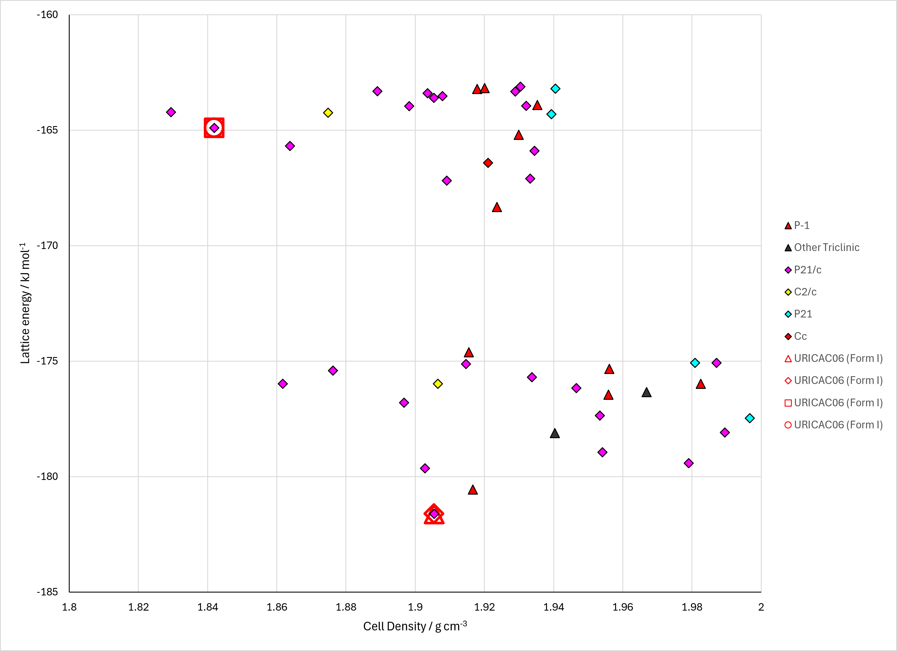
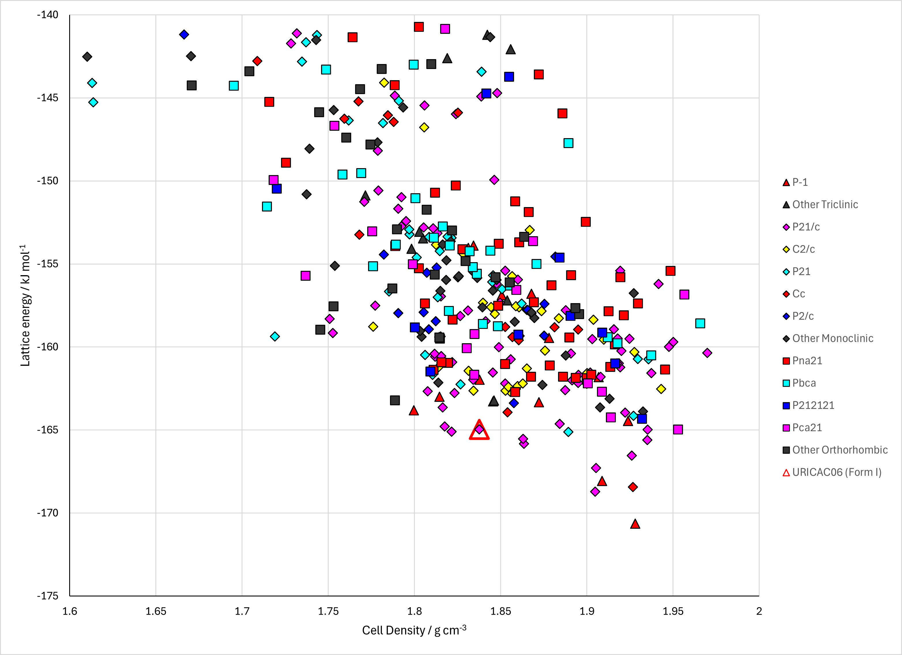
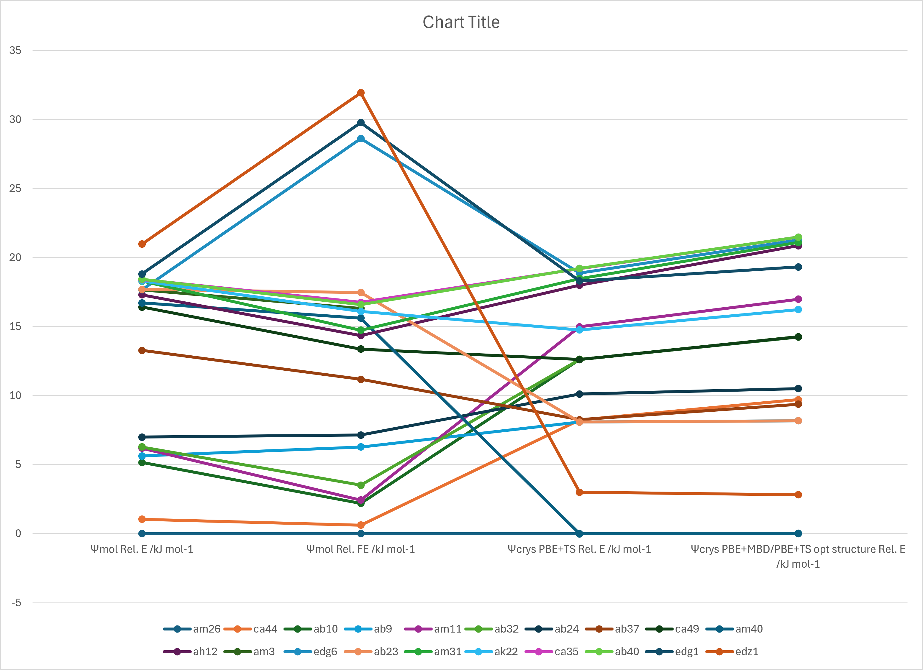
Figure 2. Lattice Energy Landscapes of Uric Acid. Energy models A1 and B1. Line chart comparing DFT+D energy models.
CSD structures (CSD version 6.01)
Table 1. Crystallographic information and experimental information of CSD entries for Uric Acid. Different polymorphs, as ascertained by structure matching of 20 molecule coordination sphere in Mercury, are coloured differently. Reference polymorphs are outlined in bold.
| REFCODE | Spacegroup | Z' | a / Å | b / Å | c / Å | α / ° | β / ° | γ / ° | density / g cm-3 | Form | R factor | T / K | Year | Comment / reference | REFCODE |
|---|
| URICAC | P21/a | 1.0 | 14.464 | 7.40300 | 6.208 | 90.0 | 65.1 | 90.0 | 1.8519 | None | 6.600 | None | 1966 | 10.1107/S0365110X66000914 | URICAC |
| URICAC01 | P21/n | 1.0 | 6.22880 | 7.3113 | 13.1274 | 90.0 | 90.632 | 90.0 | 1.8678 | None | 5.56 | 173 K | 2019 | 10.3390/min9060373 | URICAC01 |
| URICAC02 | P21/n | 1.0 | 6.2182 | 7.35300 | 13.1232 | 90.0 | 90.519 | 90.0 | 1.8610 | None | 5.26 | 213 K | 2019 | 10.3390/min9060373 | URICAC02 |
| URICAC03 | P21/n | 1.0 | 6.211 | 7.3857 | 13.1213 | 90.0 | 90.447 | 90.0 | 1.8551 | None | 4.81 | 243 K | 2019 | 10.3390/min9060373 | URICAC03 |
| URICAC04 | P21/n | 1.0 | 6.20250 | 7.41630 | 13.1147 | 90.0 | 90.378 | 90.0 | 1.8509 | None | 4.62 | 273 K | 2019 | 10.3390/min9060373 | URICAC04 |
| URICAC05 | P21/n | 1.0 | 6.1963 | 7.4512 | 13.1114 | 90.0 | 90.282 | 90.0 | 1.8445 | None | 4.2 | 303 K | 2019 | 10.3390/min9060373 | URICAC05 |
| URICAC06 | P21/n | 1.0 | 6.1898 | 7.48570 | 13.1103 | 90.0 | 90.217000 | 90.0 | 1.8381 | None | 3.65 | 333 K | 2019 | 10.3390/min9060373 | URICAC06 |
| URICAC07 | P21/n | 1.0 | 6.23520 | 7.27560 | 13.1328 | 90.0 | 90.710000 | 90.0 | 1.8743 | None | 5.5 | 130 K | 2019 | 10.3390/min9060373 | URICAC07 |
| URICAC08 | P21/n | 1.0 | 6.2302 | 7.23680 | 13.113 | 90.0 | 90.850000 | 90.0 | 1.8888 | None | 6.75 | 120 K | 2005 | None | URICAC08 |
|